Pages

Monday, May 6, 2013

VRM: The Confidenti​al Case-files of GlaxoSmith​Kline – Cover-up, Deferral & Denial of Responsibi​lity for Vaccine-re​lated Premature Deaths

The Vaccine Industry measures human life in terms of the percentages of profits to be gained. They achieve this end by routinely skewing preliminary vaccine trial data; subsequently burying or denying reports of vaccine-derived neurological trauma & neuro-developmental disorders affiliated directly with the shots.  
Vaccine Manufacturers are legally immune from prosecution for infant deaths in cases involving potential vaccine related trauma. So as a result, mainstream Pediatricians, who admittedly receive financial kickbacks for pushing Big Pharma products on their patients, (a clear conflict-of-interest type relationship which is fostered beginning in Medical school through seminars, promotions, grants & other lucrative incentives; an alliance which extends to private/public clinics & hospitals through major donations ie. supplying medical equipment, upgrading infrastructure, passing off free samples etc; including highly coveted medical research positions in the field) are subsequently hamstrung in their capacity and/or willingness to attribute any association to these shots, despite often substantial evidence pointing to an immediate causal link.
The following confidential case-files pertaining to the GlaxoSmithKline vaccine, Infanrix hexa™, were recently uncovered; a glaring exposé into Vaccine Industry malfeasance, which reveal the systemic cover-up, deferral & denial of responsibility for vaccine-related premature deaths (among infants) by vaccine manufacturing giant GlaxoSmithKline. Bare in mind, this startling evidence only represents the tip of the iceberg, given the fact that a mere 10% of vaccine related adverse reports are ever submitted by families – what is termed ‘underreporting’.
The average age of deaths among infants reported by the attending Pediatricians range from 7-weeks-old to 4-months-old. All the infants received multiple injections of Infanrix hexa™, averaging 2-3 doses. All reported deaths occurred within (2-11) days of receiving multiple vaccines, attributed to the “preferred term” Sudden infant death syndrome (SIDS), with subsequent autopsies found to be either “inconclusive” or showing “no obvious cause of death”.
Adverse effects compiled include the following: Cardiac arrest, Convulsion, Hypokinesia,  Depressed level of consciousness, Mouth haemorrhage, Nasopharyngitis, Asphyxia, Apnoeic attack, Pallor, Oxygen saturation decreased, Heart rate decreased, Sepsis, Viral infection, Pyrexia, Loss of consciousness, Cyanosis, Metabolic disorder, Ataxia, Balance disorder, Diplopia, Strabismus, Nervous system disorder, Adverse drug reaction, Death.
Excerpts from case files -
‘Two days after vaccinations, the subject died in bed. ‘
‘Less than one day after vaccination, the subject experienced convulsions.’
‘Four days after vaccination with Infanrix hexa™ and Prevenar, the subject was brought to day care centre. At 12:00, the subject was nonresponsive and had blood in his mouth.’
‘…sudden infant death in a 7-week-old female four days after vaccination with unspecified doses of Rotarix and Infanrix hexa™…subject experienced suffocation during sleep.’
‘Approximately nine days post-vaccinations, the subject was found lifeless in bed in supine position covered by a cushion/pillow. An emergency physician was only able to certify death.’
‘Approximately 3 days after vaccinations in the morning the subject experienced apnoea. When the emergency care team arrived the subject was unconscious. Cardiac arrest with apnoea and asystole was diagnosed. Resuscitation was unsuccessful.’
‘Five days after vaccinations, the subject experienced ataxia, instability and diplopia (described as strabismus). The physician suspected a possible neurological alteration.’
‘Eleven days after vaccination the subject experienced death NOS. The subject experienced adverse drug reaction and was found dead in her bed after her afternoon nap. The subject had no concomitant medication and no relevant medical history.’
‘This case was reported by the German regulatory authority and described the occurrence of death in a 12-week-old male who was vaccinated with unspecified doses of Infanrix hexa™ and Prevenar on 9 January 2006. Approximately 11 days post-vaccination, the subject died.’
‘One day after vaccinations, the subject was hospitalised to a paediatric intensive care unit and reanimated, but died from unknown cause.’
‘Next morning after vaccinations, the subject was normally drinking and was put in bed. Approximately two to three hours later, the subject was found lifeless in bed in supine position.’
‘This case was reported by a physician and described the occurrence of possible SIDS in a 3-month-old male who was vaccinated with unspecified doses of Infanrix hexa™ and Prevenar on 29 December 2009. Later on in the evening, the subject was found dead under unknown circumstances in bed.’
‘…sudden infant death syndrome in a 2-month-old male who was vaccinated with unspecified doses of Infanrix hexa™, RotaTeq and Prevenar on 27 April 2010…Twelve hours after vaccination, the subject went dusky and experienced apnoea attack, reduced oxygen saturation and decreased heart rate…Three days after discharge, the subject had another episode of apnoea and could not be resuscitated. The subject died from sudden infant death syndrome 5 days after vaccination.’
‘…occurrence of cardiovascular arrest in a 3-month-old male who was vaccinated with the 2nd dose of Infanrix hexa™ and Prevenar on 29 September 2009…Approximately three days post-vaccination, subject was found unconscious and the subject’s body was blue (cyanosis). Upon arrival of an emergency physician the pupils were medium wide, no pulse could be determined and oxygen saturation could not be measured. The subject was intubated and cardiopulmonary resuscitation was started. Under ongoing resuscitation the subject was transferred to a hospital. In hospital the subject was treated with adrenaline and atropine. Echocardiography and ECG both showed no detectable heart reaction. Body temperature, taken in the ear, was 39.4 degC. Resuscitation was without success and was stopped.’

Confidential
‘The 2080 reports were received from 41 countries, mainly France (645 reports, 31%), Italy (602 reports, 28.9%) and Germany (339 reports, 16.3%). Based on the initial reporting source, 783 cases were received from healthcare professionals and 1297 cases by non-healthcare professionals (consumers, regulatory authorities, representatives, literature, other).’ – Page 709
‘This analysis shows that the number of sudden death cases reported within 19 days of Infanrix hexa™ vaccination is below the number of cases expected for this time period in children under 2 years of age except when death occurred during the first three days after vaccination in the second year of life where the observed death number is almost equal to the number expected. The Company will continue to monitor these cases and their reporting frequencies.’ – Page 783
Case B0601431A – MedDRA Preferred Term (Sudden infant death syndrome): This case was reported by a healthcare professional and described the occurrence of cot death in a 3-month-old female who was vaccinated with the 2nd doses of Infanrix hexa™ and Prevenar on 21 October 2009. Two days after vaccinations, the subject died in bed. The parents found the baby lying on the belly. Autopsy did not reveal any cause of death.
Company comment: Suspected case of SIDS, autopsy did not reveal clear cause of death. No details on medical history available. The subject received concomitant vaccination with Prevenar.
Case B0605003A – MedDRA Preferred Terms (Cardiac arrest, Convulsion, Hypokinesia): This case was reported by the Italian regulatory authority and described the occurrence of cardiac arrest in a 2-month-old female who was vaccinated with an unspecified dose of Infanrix hexa™ on 10 August 2009. Less than one day after vaccination, the subject experienced convulsions. The subject was hospitalised from 14 August until 19 August 2009. At the time of reporting, the event was resolved with sequelae. Last convulsion episode was on 18 October 2009. The baby showed a regular growth but a light motor retardation in respect of the age. Her weight was 7.10 kg. Diagnostic tests as karyotype, ultrasonography, computerized axial tomography and nuclear magnetic resonance were negative. She was treated with Luminalette. According to the follow up information received on 01 June 2010, the subject died due to a cardiac arrest at an unspecified time after vaccination.
Company comment: (Case of Sudden Unexpected Death in Infancy (SUDI). The subject had a history of convulsions since 2-months of age, which started less than one day after vaccination with Infanrix hexa™. The final diagnosis was not reported, however, the child received anticonvulsive treatment. Cause of death was reported as cardiac arrest, but circumstances of death were not available. It was unknown whether an autopsy was performed.
Case B0608494A – MedDRA Preferred Terms (Sudden infant death syndrome, Depressed level of consciousness, Mouth haemorrhage, Nasopharyngitis): This case was reported by a healthcare professional and described the occurrence of cot death in a 14-week-old male who was vaccinated with the 2nd dose of Infanrix hexa™ and Prevenar on 12 November 2009. The child was born at term and weighed 4120 g. The child had a history of viral infection before vaccination with the 1st dose of Infanrix hexa™ and Prevenar. In the beginning of November, 2 weeks before death, the subject had a common cold. The subject did not experience any adverse events after vaccination. Four days after vaccination with Infanrix hexa™ and Prevenar, the subject was brought to day care centre. He had no fever.
He burped well after being fed and was put into bed at 9:25 lying on the abdomen (with permission of the mother) and he was being checked every 20 minutes. At 12:00, the subject was nonresponsive and had blood in his mouth. Reanimation was started immediately and the the child was admitted to hospital. The child died on 16 November 2009 from sudden infant death syndrome. An autopsy was performed and did not reveal any cause of death found in autopsy or on toxicological investigation.
Company comment: Possible case of SIDS. The subject had viral infections as medical history. No adverse events were reported after vaccinations. The subject was placed in prone position into bed. No clear cause of death was found on the autopsy.
Case B0639243A – MedDRA Preferred Terms: (Sudden infant death syndrome, Asphyxia): This case was reported by a physician and described the occurrence of sudden infant death in a 7-week-old female four days after vaccination with unspecified doses of Rotarix and Infanrix hexa™. The reporter informed that the subject experienced suffocation during sleep.
Company comment: Case of SUDI or suspected SIDS. No medical history and circumstances of death were reported. The subject, according to the reporter, experienced asphyxia during sleep. It was unknown whether an autopsy was performed.
Case B0657890A – MedDRA Preferred Terms (Sudden infant death syndrome, Apnoeic attack, Pallor, Oxygen saturation decreased, Heart rate decreased): This case was reported by a healthcare professional and described the occurrence of sudden infant death syndrome in a 2-month-old male who was vaccinated with unspecified doses of Infanrix hexa™, RotaTeq and Prevenar on 27 April 2010. Concurrent medical condition included premature birth at 26 weeks of gestation. Twelve hours after vaccination, the subject went dusky and experienced apnoea attack, reduced oxygen saturation and decreased heart rate. The subject was hospitalised. Relevant test results included: heart rate more than 100 bpm, pO2 over 94 %, normal cranial ultrasound and ophthalmological examination. The subject was treated with mechanical ventilation, stayed under observation for 48 hours and was discharged. Three days after discharge, the subject had another episode of apnoea and could not be resuscitated. The subject died from sudden infant death syndrome 5 days after vaccination.
Company comment: Case of SUDI. The subject died due to apnoea attack, likely related to his severe prematurity. It was unknown whether an autopsy was performed.
Case D0064259A – MedDRA Preferred Terms (Cardiac arrest, Sudden infant death syndrome, Sepsis, Viral infection, Resuscitation, Pyrexia, Loss of consciousness, Cyanosis): This case was reported by the German regulatory authority and described the occurrence of cardiovascular arrest in a 3-month-old male who was vaccinated with the 2nd dose of Infanrix hexa™ and Prevenar on 29 September 2009. The subject’s parents have separated about two weeks prior to the events. The subject was cared for by the father with the help of his sister-in-law and mother-in-law. The subject did not experience any adverse event between date of vaccination and date of death. Approximately three days post-vaccination, in the morning around 07:30 the subject appeared normal. About half an hour later, at around 08:00, the subject was supposed to be fed with a bottle. The subject was found unconscious and the subject’s body was blue (cyanosis). Upon arrival of an emergency physician the pupils were medium wide, no pulse could be determined and oxygen saturation could not be measured. The subject was intubated and cardiopulmonary resuscitation was started. Under ongoing resuscitation the subject was transferred to a hospital. In hospital the subject was treated with adrenaline and atropine. Echocardiography and ECG both showed no detectable heart reaction. Body temperature, taken in the ear, was 39.4 degC. Resuscitation was without success and was stopped. An autopsy was performed, but results were not conclusive. According to autopsy both SIDS and viral infection were possible causes of death. External force and shaken baby syndrome were excluded by autopsy.
Company comment: Case of SUDI. The subject died due to cardiac arrest 3 days after multiple vaccinations. The autopsy results were inconclusive and considered SIDS and viral infection as possible causes of death.
Case D0064689A – MedDRA Preferred Term (Sudden infant death syndrome): This case was reported by the German regulatory authority and described the occurrence of SIDS in 3-month-old male subject who was vaccinated with unspecified doses of Synflorix and Infanrix hexa™ on 04 November 2009. The subject has no underlying or concurrent medical conditions or other risk factors. The subject has received previous vaccination with Synflorix™ and Infanrix hexa™. It was unknown how previous vaccinations were tolerated. Approximately nine days post-vaccinations, the subject was found lifeless in bed in supine position covered by a cushion/pillow. An emergency physician was only able to certify death. Police reported that the children’s room was severely overheated and in the whole apartment people had been smoking. Autopsy was performed and showed age-corresponding state of development and very good state of care. Multipe punctual haemorrhages up to the size of a pinhead were found under the thymus capsule, subepicardial and on the surface of the lungs. Distinct disorder of blood distribution was seen in the lungs as well as increased fluid and blood content in the lungs and foam in the respiratory tract (pulmonary edema). Neither signs of external force by a third party nor signs of shaken baby syndrome have been detected.
No signs of organic malformation have been detected. The cause of death could not be unambiguously determined. Punctual haemorrhages under the thymus capsule, subepicardial and on the surface of the lungs were normally seen within the scope of SIDS and therefore the autopsy performing physicians considered SIDS. Possible risk factors associated with SIDS included coverage with a pillow, severely overheating, not feeding with breast milk and passive smoking. Furthermore autopsy showed increased water retention of the lungs as well as distinct disorder of blood distribution within the lungs, which could be signs of a beginning pulmonary infection. Microbiological examinations, performed on 20 November 2009, showed solitary St. aureus in both pulmonary swabs and a single St. aureus colony in the spleen swab as potential infectious agent, but this bacterium was also known as normal bacterial flora of the upper respiratory tract. All other bacteria found belong either to physiological intestinal flora or were normal parts of the throat and skin flora. Therefore infectious events could be excluded with some probability.
Company comment: Case of SUDI. The subject died 9 days after multiple vaccinations. The autopsy results were inconclusive and considered SIDS (in presence of numerous risk factors) and pulmonary infection as possible causes of death.
Case D0065445A – MedDRA Preferred Term (Sudden infant death syndrome): This case was reported by a physician and described the occurrence of SIDS in a 3-month-old female one day after vaccination with the 1st dose of Infanrix hexa™ and Prevenar on 09 December 2009. The subject’s development and weight gain were normal, her medical history was unsuspicious. She was breast-fed for three months. Postvaccinations the subject did not experience fever. Next morning after vaccinations, the subject was normally drinking and was put in bed. Approximately two to three hours later, the subject was found lifeless in bed in supine position. The subject died on 10 December 2009 from SIDS. An autopsy was performed, but no results were available.
Company comment: Suspected case of SIDS. The subject died 1 day after multiple vaccinations. Subject’s medical history was unsuspicious, no adverse event were reported after vaccinations. Autopsy was performed, but the results were not available.
Case D0066068A – MedDRA Preferred Term (Sudden infant death syndrome): This case was reported by a physician and described the occurrence of possible SIDS in a 3-month-old male who was vaccinated with unspecified doses of Infanrix hexa™ and Prevenar on 29 December 2009. The subject had three healthy siblings. He was a healthy term baby and was breast-fed. The subject’s mother did not smoke. The subject had received the last meal, consisting of mother’s milk on 29 December 2009 in the evening. Later on in the evening, the subject was found dead under unknown circumstances in bed. An autopsy was performed. The results of autopsy were inconclusive and showed no obvious cause of death. Therefore cause of death was considered to be SIDS.
Company comment: Possible case of SIDS. The subject died less than 1 day after multiple vaccinations. Subject’s medical history included healthy term baby, without underlying condition. Autopsy did not reveal clear cause of death.
Case D0067790A – MedDRA Preferred Terms (Sudden infant death syndrome, Death, Apnoea, Cardiac arrest, Loss of consciousness, Resuscitation): This case was reported by the German regulatory authority and described the occurrence of SIDS in a 9-week-old male who was vaccinated with Infanrix hexa™ and Prevenar on 31 March 2010. Complication during pregnancy included cranial haemorrhage of the mother due to cerebral artery aneurysm in the 19th week of gestation. The subject was born in the 33rd week of pregnancy by Caesarean section. At that time the subject was immature with a birth weight of 1805g with mild respiratory distress syndrome. Postnatal the subject showed good adaptation, but chest X-ray, performed on 28 January 2010, showed mixed picture of mild neonatal respiratory distress syndrome and wet lung. Repeated sonography and neonatal screening were normal. Concurrent medications included colecalciferol and iron salt. For the third child health check, performed on 04 March 2010, the subject showed normal development concerning weight, length and head circumference.
The subject showed no pathologic findings except mild hydrocele. However, according to percentile curve of the WHO the subject was in reduced nutritional condition with a weight of 3700g and a height of 55cm. Approximately 3 days after vaccinations in the morning the subject experienced apnoea. When the emergency care team arrived the subject was unconscious. Cardiac arrest with apnoea and asystole was diagnosed. Resuscitation was unsuccessful. Concurrent medical conditions included old contusion and hematoma on right side of chest. An autopsy was performed and macroscopically, did not reveal unambiguous cause of death. All autopsy findings were suggestive for SIDS. The findings not consistent with SIDS (skin fissures in the corner of mouth, ecchymoses in area of central chest wall, hemorrhage in capsule of adrenal gland and kidney) can be explained with plausibility by long and continuous resuscitation. Further toxicological examination could not identify the cause of death. According to the report on the histological examination, results largely confirmed the findings of the autopsy.
Besides unspecific signs of death, punctuate haemorrhage of the organs’ connective tissue coatings and pulmonary emphysema were considered the essential findings. Acute emphysema could be interpreted as evidence of suffocation. It was concluded that definite cause of death could be identified, neither in histological examinations nor in toxicological tests. It was discussed that the toxicological tests covered a certain spectrum of substances only and would miss some rare and exceptional substances. Because of the combination of pulmonary emphysema and the fissures at the left corner of the mouth, which had been observed during the autopsy, death due to suffocation following violent obstruction of respiratory orifices could not be excluded. Likewise it could not be excluded that these findings were caused during the reanimation procedures.
Company comment: Case of SUDI. Preterm subject, with respiratory distress in medical history, died approximately 3 days after multiple vaccinations. Autopsy did not reveal clear cause of death. Majority of findings were within SIDS, however violent obstruction of respiratory tract could not be excluded.
Case B0661542A – MedDRA Preferred Terms (Metabolic disorder, Ataxia, Balance disorder, Diplopia, Strabismus, Nervous system disorder): This case was reported by a physician and described the occurrence of ataxia in a 6-month-old male who was vaccinated with the 3rd doses of Infanrix hexa™ and Prevenar in March 2010. The subject’s medical history included episodes of shaking head, arms and legs several times a day, which occurred at the age of 5 months. Five days after vaccinations, the subject experienced ataxia, instability and diplopia (described as strabismus). The physician suspected a possible neurological alteration. The subject was hospitalized and some relevant tests (NMR, ECG, CSF, other unspecified laboratory tests, nasopharyngeal exudates) were performed and showed normal results. Catecholamine and muscular biopsy results were pending. The ataxia remained until the age of 9 months. The shaking moves have repeated in some occasions. The subject was hospitalized in an intensive care due to a possible aspiration from 16 to 24 June 2010. He underwent EEG in July 2010 and it showed 3 lesions compatible with metabolic disorder. The final diagnosis was a possible metabolic disease with very limited clinical picture. According to the follow-up information received in August 2010, the subject died in July 2010 due to a possible metabolic disorder of a mitochondrial origin.
Company comment: The subject died due to a possible metabolic disorder of a mitochondrial origin several months after 3d vaccination with Infanrix hexa™ and Prevenar. It was unknown whether an autopsy was performed.
Case B0599802A- MedDRA Preferred Term (Death, Adverse drug reaction): This case was reported by a healthcare professional and described the occurrence of death NOS (not otherwise specified) in a 4-month-old female who was vaccinated with the 3rd dose of Infanrix hexa™ and Prevenar on 15 October 2009. Eleven days after vaccination the subject experienced death NOS. The subject experienced adverse drug reaction and was found dead in her bed after her afternoon nap. The subject had no concomitant medication and no relevant medical history. The subject was transferred to hospital. Hospital report was pending. No autopsy was performed. Follow-up information has been requested.
Case D0063296A – MedDRA Preferred Term (Death): This case was reported by the German regulatory authority and described the occurrence of death in a 12-week-old male who was vaccinated with unspecified doses of Infanrix hexa™ and Prevenar on 9 January 2006. The subject‟s mother suffered from epilepsy. The subject was exposed in utero to levetiracetam during about the first three months of pregnancy. The rest of pregnancy and birth was inconspicuous, except for fracture of a clavicle. Concurrent medical conditions included agitation and crying abnormal (whiny baby). Approximately 11 days post-vaccination, the subject died. The cause of death was not further specified. It was unknown whether an autopsy was performed.
Case D0069211A – MedDRA Preferred Term (Death): This case was reported by a physician and described the occurrence of unspecified death in a child of unspecified gender who was vaccinated on an unspecified date with an unspecified 6-valent vaccine and unspecified pneumococcal vaccine (manufacturers unspecified). One day after vaccinations, the subject was hospitalised to a paediatric intensive care unit and reanimated, but died from unknown cause. An autopsy was performed. Follow-up information has been requested.
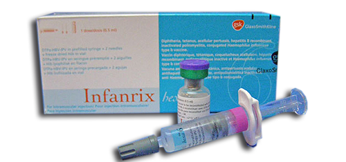
infanrix-1
Package Insert – Infanrix hexa™ (Combined Diptheria, Tetanus and Acellular Pertusis, Hepatits B inhanced Inactivated Poliomyelitis Haemophilus influenzae type B vaccine)
Infanrix hexa™ contains the following antigens adsorbed onto aluminium salts: diphtheria toxoid, tetanus toxoid, three purified pertussis antigens pertussis toxoid (PT), filamentous haemagglutinin (FHA) and pertactin (PRN; 69 kiloDalton outer membrane protein) , the purified major surface antigen (HBsAg) of the hepatitis B virus (HBV) and purified polyribosyl-ribitol-phosphate capsular polysaccharide (PRP) of Haemophilus influenzae type b (Hib), covalently bound to tetanus toxoid. It also contains three types of inactivated polio viruses (type 1: Mahoney strain; type 2: MEF-1 strain; type 3: Saukett strain).
The tetanus and diphtheria toxoids are obtained by formaldehyde treatment of purified Corynebacterium diphtheriae and Clostridium tetani toxins. The acellular pertussis vaccine components are obtained by extraction and purification from phase I Bordetella pertussis cultures, followed by irreversible detoxification of the pertussis toxin by glutaraldehyde and formaldehyde treatment, and formaldehyde treatment of FHA and PRN. The diphtheria toxoid, tetanus toxoid and acellular pertussis components are adsorbed onto aluminium salts.
The surface antigen of the HBV is produced by culture of genetically-engineered yeast cells (Saccharomyces cerevisiae) which carry the gene coding for the major surface antigen of the HBV. This HBsAg expressed in yeast cells is purified by several physicochemical steps. The HBsAg assembles spontaneously, in the absence of chemical treatment, into spherical particles of 20 nm in average diameter containing nonglycosylated HBsAg polypeptide and a lipid matrix consisting mainly of phospholipids. Extensive tests have demonstrated that these particles display the characteristic properties of the natural HBsAg.
The three polioviruses are cultivated on a continuous VERO cell line, purified and inactivated with formaldehyde.
The Hib polysaccharide is prepared from Hib, strain 20,752 and after activation with cyanogen bromide and derivatisation with an adipic hydrazide spacer is coupled to tetanus toxoid via carbodiimide condensation. After purification the conjugate is adsorbed on aluminium salt, and then lyophilised in the presence of lactose as stabiliser. Infanrix hexa™ meets the World Health Organisation requirements for manufacture of biological substances, of diphtheria, tetanus, pertussis and combined vaccines, of hepatitis B vaccines made by recombinant DNA techniques, of inactivated poliomyelitis vaccines and of Hib conjugate vaccines.
Infanrix hexa™ is indicated for primary and booster immunisation against diphtheria, tetanus, pertussis, hepatitis B, poliomyelitis and Haemophilus influenzae type b in infants from the age of 6 weeks and may be given to infants who received a first dose of hepatitis B vaccine at birth.
The primary vaccination schedule (such as 2, 3, 4 months; 3, 4, 5 months; 2, 4, 6 months; 3, 5 and 11 or 12 months; 6, 10, 14 weeks) consists of three doses of 0.5 ml. An interval of at least one month should be respected between doses. If it is intended to administer Infanrix hexa™ according to the EPI schedule (Expanded Program on Immunisation; 6, 10, 14 weeks of age), then the vaccinee must receive a dose of hepatitis B vaccine at birth.
A 0.5 ml dose of the vaccine contains not less than 30 IU of adsorbed diphtheria toxoid, not less than 40 IU of adsorbed tetanus toxoid, 25 mcg of adsorbed PT, 25 mcg of adsorbed FHA, 8 mcg of adsorbed pertactin, 10 mcg of adsorbed recombinant HBsAg protein, 40 D-antigen units of type 1 (Mahoney), 8 D-antigen units of type 2 (MEF-1) and 32 D-antigen units of type 3 (Saukett) of the polio virus. It also contains 10 mcg of adsorbed purified capsular polysaccharide of Hib (PRP) covalently bound to 20-40 mcg tetanus toxoid (T).
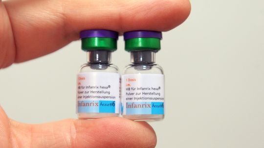
infanrix-hexa
GlaxoSmith​Kline (GSK) Confidential to regulatory agencies file: http://ddata.over-blog.com/3/27/09/71/2012-2013/confid.pdf
Related Articles:
VRM: GlaxoSmithKline Implicated In Systemic Clinical Drug Trial Deception & Insider Fraud http://vaccineresistancemovement.org/?p=6838
VRM: Mandatory Vaccinatio​ns – How They Will Be Implemente​d http://vaccineresistancemovement.org/?p=11806
VRM: The Autism Report http://vaccineresistancemovement.org/?p=10185
VRM: Worldwide Autism Study Direct link to study: http://study.vaccineresistancemovement.org/
VRM: The Problem With Vaccines Part 1 http://vaccineresistancemovement.org/?p=488
VRM: Vaccine Clinic – A Concise Compendium To The Problem With Vaccines http://vaccineresistancemovement.org/?p=6278
VRM: The Problem With Vaccines Part 2 – Synergistic Effect of Heavy Metal Toxicity On The Body http://vaccineresistancemovement.org/?p=6097
VRM: The Problem With Vaccines Part 3 – Synthetic Genomics & The Death Of Natural Immunity http://vaccineresistancemovement.org/?p=6880
VRM: A Concise Compendium To The Problem With Vaccines Part 3 – Synthetic Genomics & The Death Of Natural Immunity http://vaccineresistancemovement.org/?p=7283
VRM: The Problem With Vaccines Part 4 – Primary Aspects of Vaccine Toxicity Affecting Body http://vaccineresistancemovement.org/?p=8787
VRM: The Problem With Vaccines Part 5A – Detoxification & Restoration of the Body http://vaccineresistancemovement.org/?p=8836
VRM: The Problem With Vaccines Part 5B – Detoxification & Restoration of the Body http://vaccineresistancemovement.org/?p=8847
VRM: PCV Vaccine Exposed – Breeding Ground For Staphylococcus Aureus http://vaccineresistancemovement.org/?p=9431
VRM: Pandemic Preparedness & The Dark Agenda Ahead http://vaccineresistancemovement.org/?p=9460
VRM: The Flu Report http://vaccineresistancemovement.org/?p=9226
VRM: Vaccine Ingredients http://vaccineresistancemovement.org/?p=979
VRM: Safe Alternatives to Vaccines http://vaccineresistancemovement.org/?p=662%EF%BB%BF
VRM: Family Charts Gradual Decline Of Daughter http://vaccineresistancemovement.org/?p=3156
VRM: Health Matters Part 1 http://vaccineresistancemovement.org/?p=6719
VRM: Health Matters Part 2 http://vaccineresistancemovement.org/?p=6746%EF%BB%BF
VRM: Alternative Cancer Cures That Work http://vaccineresistancemovement.org/?p=3729
VRM: Pregnancy Tips http://vaccineresistancemovement.org/?p=3270
VRM: H1N1 Shot Reactions – Miscarriages http://vaccineresistancemovement.org/?p=943
VRM: The Vanishing Sperm Count http://vaccineresistancemovement.org/?p=4639
VRM: H1N1 Vaccine Surplus From 2009 Reveals Growing Distrust of Gov’t & WHO http://vaccineresistancemovement.org/?p=4969
VRM: Flu Death Statistics – WHO & The Big Lie http://vaccineresistancemovement.org/?p=784
VRM: Vaccine Industry Deception, Propaganda & Media Collusion http://vaccineresistancemovement.org/?p=197
VRM: Birth of Medical & Scientific Dictatorship – Future Scenarios http://vaccineresistancemovement.org/?p=997
VRM: H1N1 Bio-weaponry Incorporated http://vaccineresistancemovement.org/?p=884
VRM: Aids & The WHO Connection – Criminal Intent http://vaccineresistancemovement.org/?p=1749
VRM: Morgellons Syndrome & Chemtrails http://vaccineresistancemovement.org/?p=839
VRM: Council On Foreign Relations 10/16/09- Major Influence on Government Vaccine Policy http://vaccineresistancemovement.org/?p=1880
VRM: Closed Door CDC Meeting Reveals Industry Cover-up Of Heavy Metal Toxicity In Vaccines http://vaccineresistancemovement.org/?p=5935
VRM: The Rockefeller Foundation Drafts A Post-Pandemic Scenario http://vaccineresistancemovement.org/?p=5102
VRM: World Health Organization & Vaccine Manufacturers Implicated In Massive H1N1 Financial Scam Involving Kickbacks & Cover-ups http://vaccineresistancemovement.org/?p=4610
VRM Live – 01/28/11: Vaccine Resistance Movement founder Joel    Lord  discusses Synthetic Genomics, cloned cell vaccine technology    & the  death of natural immunity, gutter journalism & Dr.    Wakefield’s  imminent vindication with ‘Truth to Power’ host Paul    Mabelis. http://www.blogtalkradio.com/empradio/2011/01/28/truth-to-power-thursday
VRM Live - 11/04/10: Vaccine     Resistance Movement founder Joel Lord lays out the whole vaccine    process  with Paul Mabelis; including heavy metal toxicity, synergy,     pregnancy issues & the basic principles of natural health at risk. http://www.blogtalkradio.com/show.aspx?userurl=empradio&year=2010&month=11&day=05&url=truth-to-power-thursday
VRM Live - 09/24/10:    Vaccine  Resistance Movement Founder Joel Lord & activist/radio    host Jesse  Calhoun lay it all out tonite. Topics include the VRM    Worldwide Autism Study, Scientific/Medical  dictatorship, Natural Rights & Vaccine Industry fraud exposed. Special thanks to host Paul Mabelis. http://www.blogtalkradio.com/empradio/2010/09/24/truth-to-power-thursday
If you appreciate the efforts to bring this information   forward  do consider making a donation. Any amount, no matter how small   will  help enable me to carry on this invaluable research. See Paypal   link  on the VRM website (click on ‘Donate’ tab in upper right corner). Thank you all.

Child Abuse Hotline Ad Uses Photographic Trick That Makes It Visible Only To Children

Saturday, May 4, 2013

Elder Abuse and Neglect

What is elder abuse?


As elders become more physically frail, they’re less able to stand up to bullying and or fight back if attacked. They may not see or hear as well or think as clearly as they used to, leaving openings for unscrupulous people to take advantage of them. Mental or physical ailments may make them more trying companions for the people who live with them.
Many seniors around the world are being abused: harmed in some substantial way often by people who are directly responsible for their care.
In the U.S. alone, more than half a million reports of abuse against elderly Americans reach authorities every year, and millions more cases go unreported.


Where does elder abuse take place?

 Elder abuse tends to take place where the senior lives: most often in the home where abusers are apt to be adult children; other family members such as grandchildren; or spouses/partners of elders. Institutional settings, especially long-term care facilities, can also be sources of elder abuse. 

The different types of elder abuse

Abuse of elders takes many different forms, some involving intimidation or threats against the elderly, some involving neglect, and others involving financial chicanery. The most common are defined below.

Physical abuse

Physical elder abuse is non-accidental use of force against an elderly person that results in physical pain, injury, or impairment. Such abuse includes not only physical assaults such as hitting or shoving but the inappropriate use of drugs, restraints, or confinement.

Emotional abuse

In emotional or psychological senior abuse, people speak to or treat elderly persons in ways that cause emotional pain or distress.
Verbal forms of emotional elder abuse include
  • Intimidation through yelling or threats
  • Humiliation and ridicule
  • Habitual blaming or scapegoating
Nonverbal psychological elder abuse can take the form of
  • Ignoring the elderly person
  • Isolating an elder from friends or activities
  • Terrorizing or menacing the elderly person

Sexual abuse

Sexual elder abuse is contact with an elderly person without the elder’s consent. Such contact can involve physical sex acts, but activities such as showing an elderly person pornographic material, forcing the person to watch sex acts, or forcing the elder to undress are also considered sexual elder abuse.

Neglect or abandonment by caregivers

Elder neglect, failure to fulfill a caretaking obligation, constitutes more than half of all reported cases of elder abuse. It can be active (intentional) or passive (unintentional, based on factors such as ignorance or denial that an elderly charge needs as much care as he or she does).

Financial exploitation

This involves unauthorized use of an elderly person’s funds or property, either by a caregiver or an outside scam artist.
An unscrupulous caregiver might
  • Misuse an elder’s personal checks, credit cards, or accounts
  • Steal cash, income checks, or household goods
  • Forge the elder’s signature
  • Engage in identity theft
Typical rackets that target elders include
  • Announcements of a “prize” that the elderly person has won but must pay money to claim
  • Phony charities
  • Investment fraud

Healthcare fraud and abuse

Carried out by unethical doctors, nurses, hospital personnel, and other professional care providers, examples of healthcare fraud and abuse regarding elders include
  • Not providing healthcare, but charging for it
  • Overcharging or double-billing for medical care or services
  • Getting kickbacks for referrals to other providers or for prescribing certain drugs
  • Overmedicating or undermedicating
  • Recommending fraudulent remedies for illnesses or other medical conditions
  • Medicaid fraud

Signs and symptoms of elder abuse

At first, you might not recognize or take seriously signs of elder abuse. They may appear to be symptoms of dementia or signs of the elderly person’s frailty — or caregivers may explain them to you that way. In fact, many of the signs and symptoms of elder abuse do overlap with symptoms of mental deterioration, but that doesn’t mean you should dismiss them on the caregiver’s say-so.

General signs of abuse

The following are warning signs of some kind of elder abuse:
  • Frequent arguments or tension between the caregiver and the elderly person
  • Changes in personality or behavior in the elder
If you suspect elderly abuse, but aren't sure, look for clusters of the following physical and behavioral signs.

Physical abuse

  • Unexplained signs of injury such as bruises, welts, or scars, especially if they appear symmetrically on two side of the body
  • Broken bones, sprains, or dislocations
  • Report of drug overdose or apparent failure to take medication regularly (a prescription has more remaining than it should)
  • Broken eyeglasses or frames
  • Signs of being restrained, such as rope marks on wrists
  • Caregiver’s refusal to allow you to see the elder alone

Emotional abuse

In addition to the general signs above, indications of emotional elder abuse include:
  • Threatening, belittling, or controlling caregiver behavior that you witness
  • Behavior from the elder that mimics dementia, such as rocking, sucking, or mumbling to oneself

Sexual abuse

  • Bruises around breasts or genitals
  • Unexplained venereal disease or genital infections
  • Unexplained vaginal or anal bleeding
  • Torn, stained, or bloody underclothing

Neglect by caregivers or self-neglect

  • Unusual weight loss, malnutrition, dehydration
  • Untreated physical problems, such as bed sores
  • Unsanitary living conditions: dirt, bugs, soiled bedding and clothes
  • Being left dirty or unbathed
  • Unsuitable clothing or covering for the weather
  • Unsafe living conditions (no heat or running water; faulty electrical wiring, other fire hazards)
  • Desertion of the elder at a public place

Financial exploitation

  • Significant withdrawals from the elder’s accounts
  • Sudden changes in the elder’s financial condition
  • Items or cash missing from the senior’s household
  • Suspicious changes in wills, power of attorney, titles, and policies
  • Addition of names to the senior’s signature card
  • Unpaid bills or lack of medical care, although the elder has enough money to pay for them
  • Financial activity the senior couldn’t have done, such as an ATM withdrawal when the account holder is bedridden
  • Unnecessary services, goods, or subscriptions

Healthcare fraud and abuse

  • Duplicate billings for the same medical service or device
  • Evidence of overmedication or undermedication
  • Evidence of inadequate care when bills are paid in full
  • Problems with the care facility: poorly trained, poorly paid, or insufficient staff; crowding; inadequate responses to questions about care 

Risk factors for elder abuse

It’s difficult to take care of a senior when he or she has many different needs, and it’s difficult to be elderly when age brings with it infirmities and dependence. Both the demands of caregiving and the needs of the elder can create situations in which abuse is more likely to occur.

Risk factors among caregivers

Many nonprofessional caregivers — spouses, adult children, other relatives and friends — find taking care of an elder to be satisfying and enriching. But the responsibilities and demands of elder caregiving, which escalate as the elder’s condition deteriorates, can also be extremely stressful. The stress of elder care can lead to mental and physical health problems that make caregivers burned out, impatient, and unable to keep from lashing out against elders in their care.
Among caregivers, significant risk factors for elder abuse are:
  • Inability to cope with stress (lack of resilience)
  • Depression, which is common among caregivers
  • Lack of support from other potential caregivers
  • The caregiver’s perception that taking care of the elder is burdensome and without psychological reward
  • Substance abuse
Even caregivers in institutional settings can experience stress at levels that lead to elder abuse. Nursing home staff may be prone to elder abuse if they lack training, have too many responsibilities, are unsuited to caregiving, or work under poor conditions.

The elder’s condition and history

Several factors concerning elders themselves, while they don’t excuse abuse, influence whether they are at greater risk for abuse:
  • The intensity of an elderly person’s illness or dementia
  • Social isolation; i.e., the elder and caregiver are alone together almost all the time
  • The elder’s role, at an earlier time, as an abusive parent or spouse
  • A history of domestic violence in the home
  • The elder’s own tendency toward verbal or physical aggression
In many cases, elder abuse, though real, is unintentional. Caregivers pushed beyond their capabilities or psychological resources may not mean to yell at, strike, or ignore the needs of the elders in their care.

Reporting elder abuse

If you are an elder who is being abused, neglected, or exploited, tell at least one person. Tell your doctor, a friend, or a family member whom you trust. Other people care and can help you.
And if you see an older adult being abused or neglected, don’t hesitate to report the situation. Don’t assume that someone else will take care of it or that the person being abused is capable of getting help if he or she really needs it.
Many seniors don't report the abuse they face even if they’re able. Some fear retaliation from the abuser, while others believe that if they turn in their abusers, no one else will take care of them. When the caregivers are their children, they may be ashamed that their children are behaving abusively or blame themselves: “If I’d been a better parent when they were younger, this wouldn’t be happening.” Or they just may not want children they love to get into trouble with the law.

How do I report elder abuse?

Reporting elder abuse

As difficult as reporting elder abuse can be, it’s important for you to stand up for an older adult in need. Learn how to communicate effectively in different situations and put a stop to elder abuse and neglect. Read Reporting Elder Abuse
Every state in the U.S. has at least one toll-free elder abuse hotline or helpline for reporting elder abuse in the home, in the community, or in nursing homes and other long-term care facilities. For help in your area, see the resources and references at the end of this article or talk to your local hospital or a trusted doctor or therapist.
The first agency to respond to a report of elderly abuse, in most states, is Adult Protective Services (APS). Its role is to investigate abuse cases, intervene, and offer services and advice. Again, the power and scope of APS varies from state to state.

Preventing elder abuse and neglect

We can help reduce the incidence of elder abuse, but it’ll take more effort than we’re making now. Preventing elder abuse means doing three things:
  • Listening to seniors and their caregivers
  • Intervening when you suspect elder abuse
  • Educating others about how to recognize and report elder abuse

What you can do as a caregiver to prevent elder abuse

If you’re overwhelmed by the demands of caring for an elder, do the following:
  • Request help, from friends, relatives, or local respite care agencies, so you can take a break, if only for a couple of hours.
  • Find an adult day care program.
  • Stay healthy and get medical care for yourself when necessary.
  • Adopt stress reduction practices.
  • Seek counseling for depression, which can lead to elder abuse.
  • Find a support group for caregivers of the elderly.
  • If you’re having problems with drug or alcohol abuse, get help.
And remember, elder abuse helplines offer help for caregivers as well. Call a helpline if you think there’s a possibility you might cross the line into elder abuse.

What you can do as a concerned friend or family member

  • Watch for warning signs that might indicate elder abuse. If you suspect abuse, report it.
  • Take a look at the elder’s medications. Does the amount in the vial jive with the date of the prescription?
  • Watch for possible financial abuse. Ask the elder if you may scan bank accounts and credit card statements for unauthorized transactions.
  • Call and visit as often as you can. Help the elder consider you a trusted confidante.
  • Offer to stay with the elder so the caregiver can have a break — on a regular basis, if you can.

How you can protect yourself, as an elder, against elder abuse

  • Make sure your financial and legal affairs are in order. If they aren’t, enlist professional help to get them in order, with the assistance of a trusted friend or relative if necessary.
  • Keep in touch with family and friends and avoid becoming isolated, which increases your vulnerability to elder abuse.
  • If you are unhappy with the care you’re receiving, whether it’s in your own home or in a care facility, speak up. Tell someone you trust and ask that person to report the abuse, neglect, or substandard care to your state’s elder abuse helpline or long term care ombudsman, or make the call yourself.
Finally, if you aren’t in a position to help an elder personally, you can volunteer or donate money to the cause of educating people about elder abuse, and you can lobby to strengthen state laws and policing so that elder abuse can be investigated and prosecuted more readily. The life you save down the line may be your own.


Lotus birth

Lotus birth is the practice of leaving the umbilical cord uncut after childbirth so that the baby is left attached to his/her placenta until the cord naturally separates at the umbilicus, usually a few days after birth.[1] Lotus births are rare in Western culture.
There is no evidence that there are any benefits for the health of the baby with lotus birth. A spokesman for the Royal College of Obstetricians and Gynaecologists (RCOG) has stated, "If left for a period of time after the birth, there is a risk of infection in the placenta which can consequently spread to the baby. The placenta is particularly prone to infection as it contains blood. At the post-delivery stage, it has no circulation and is essentially dead tissue," and the RCOG strongly recommends that any baby that undergoes lotus birthing to be monitored closely for infection.


Wednesday, May 1, 2013

Posttraumatic Stress Disorder (PTSD) 

Posttraumatic Stress Disorder is a complex health condition that can develop in response to a traumatic experience – a life-threatening or extremely distressing situation that causes a person to feel intense fear, horror or a sense of helplessness. PTSD can cause severe problems at home or at work. Anyone can develop PTSD – men, women, children, young and old alike. Fortunately, PTSD is treatable.

What causes PTSD?

Virtually any trauma, defined as an event that is life-threatening or that severely compromises the physical or emotional well-being of an individual or causes intense fear, may cause PTSD. Such events often include either experiencing or witnessing a severe accident or physical injury, receiving a life-threatening medical diagnosis, being the victim of kidnapping or torture, exposure to war combat or to a natural disaster, exposure to other disaster (for example, plane crash) or terrorist attack, being the victim of rape, mugging, robbery, or assault, enduring physical, sexual, emotional, or other forms of abuse, as well as involvement in civil conflict. Although the diagnosis of PTSD currently requires that the sufferer has a history of experiencing a traumatic event as defined here, people may develop PTSD in reaction to events that may not qualify as traumatic but can be devastating life events like divorce or unemployment.

What are PTSD symptoms and signs?

The following three groups of symptom criteria are required to assign the diagnosis of PTSD:Recurrent re-experiencing of the trauma (for example, troublesome memories, flashbacks that are usually caused by reminders of the traumatic events, recurringnightmares about the trauma and/or dissociative reliving of the trauma)Avoidance to the point of having a phobia of places, people, and experiences that remind the sufferer of the trauma or a general numbing of emotional responsivenessChronic physical signs of hyperarousal, including sleepproblems, trouble concentrating, irritability, anger, poor concentration, blackouts or difficulty remembering things, increased tendency and reaction to being startled, and hypervigilance (excessive watchfulness) to threatThe emotional numbing of PTSD may present as a lack of interest in activities that used to be enjoyed (anhedonia), emotional deadness, distancing oneself from people, and/or a sense of a foreshortened future (for example, not being able to think about the future or make future plans, not believing one will live much longer). At least one re-experiencing symptom, three avoidance/numbing symptoms, and two hyperarousal symptoms must be present for at least one month and must cause significant distress or functional impairment in order for the diagnosis of PTSD to be assigned. PTSD is considered of chronic duration if it persists for three months or more.A similar disorder in terms of symptom repertoire is acute stress disorder. The major differences between the two disorders are that acute stress disorder symptoms persist from two days to four weeks, and a fewer number of traumatic symptoms are required to make the diagnosis as compared to PTSD.In children, re-experiencing the trauma may occur through repeated play that has trauma-related themes instead of or in addition to memories, and distressing dreams may have more general content rather than of the traumatic event itself. As in adults, at least one re-experiencing symptom, three avoidance/numbing symptoms, and two hyperarousal symptoms must be present for at least one month and must cause significant distress or functional impairment in order for the diagnosis of PTSD to be assigned. When symptoms have been present for less than one month, a diagnosis of acute stress disorder (ASD) can be made.Symptoms of PTSD that tend to be associated with C-PTSD include problems regulating feelings, which can result in suicidal thoughts, explosive anger, or passive aggressive behaviors; a tendency to forget the trauma or feel detached from one's life (dissociation) or body (depersonalization); persistent feelings of helplessness, shame, guilt, or being completely different from others; feeling the perpetrator of trauma is all-powerful and preoccupation with either revenge against or allegiance with the perpetrator; and severe change in those things that give the sufferer meaning, like a loss of spiritual faith or an ongoing sense of helplessness, hopelessness, or despair.


Sunday, April 28, 2013

Is Rare Blood Disorder Caused by HPV Vaccines? Ladies, please read this.

Another autoimmune disorder may be plaguing some recipients of HPV vaccines. Idiopathic Thrombocytopenia Purpura, a blood disorder that causes abnormal bleeding, is found in 7 times as many girls after HPV vaccination than any other type of vaccine—a serious indication of a connection.


Kirstie was only 12 years old when she was diagnosed with a rare disorder for which there is no known cure, Idiopathic Thrombocytopenic Purpura (ITP). This happened just a little over a month after her second injection of Gardasil. She will live with this disorder for the rest of her life. Unfortunately, she is not alone.
Gardasil and Cervarix are two of the 76 FDA approved vaccines included in the VAERS (Vaccine Adverse Event Reporting System) database. Since Gardasil was approved for use in the United States, there have been 682 reports of ITP after vaccine administration. 126 of these reports occurred after HPV vaccines. If all vaccines carried equal risk, there should only be 18 reports of Idiopathic Thrombocytopenic Purpura after HPV vaccine use. Why are reports of ITP after Gardasil and Cervarix seven times that amount?


How rare is ITP?
When disease incidence rates are quoted, it is almost always in the context of number of cases per 100,000 people. This is not the case with ITP.
According to the Medscape reference library, the incidence rates for ITP are as follows:
  • United States – adults – 66 cases per 1,000,000 per year
  • United States – children – 50 cases per 1,000,000 per year
  • Denmark and England – 10 to 40 cases per 1,000,000 per year
  • Kuwait – 125 cases per 1,000,000 per year
Idiopathic thrombocytopenic purpura is such a rare disorder that a diagnosis is rendered only after all other possible conditions that could cause purpura have been eliminated. This is called a ‘diagnosis of exclusion’. How many young people are living with undiagnosed ITP after HPV vaccine administration?


What is ITP?
Idiopathic simply means ‘of unknown origin.’ Frequently the word ‘immune’ is substituted for ‘idiopathic’ when referring to ITP because of the antibodies generated to specific platelet membrane proteins that cause the person’s immune system to attack their own platelets.
Thrombocytopenia is the medical term for a low blood platelet count. Platelets (thrombocytes) are minute, disk shaped particles in the blood that promote clotting. When an injury causes a blood vessel to break, the platelets are activated causing them to become spiny. The resulting ‘spines’ allow them to stick to each other and the broken blood vessel walls to begin the clotting process. The normal amount of platelets circulating in a person’s blood ranges from 150,000 to 450,000 per micro-liter. When the blood platelet count falls below 20,000 the thrombocytopenia can cause excessive internal bleeding.
Purpura refers to purple-colored spots and patches that occur on the skin, organs, and in mucus membranes, including the lining of the mouth. Purpura is caused by internal bleeding from small blood vessels.
In short, idiopathic thrombocytopenic purpura is an autoimmune disorder caused by immunoglobulin G (IgG) auto-antibodies on the platelet surface. The number of circulating platelets is reduced due to increased destruction resulting in internal bleeding of varying degrees. ITP in children most commonly occurs following an infection, or occasionally following immunisation. Acute (sudden onset) ITP often resolves spontaneously within a few months. When ITP persists longer than 6 months without specific cause, the condition is considered chronic.


What are the symptoms of ITP?
Simply having a low platelet count does not cause symptoms. However, the bleeding that a low platelet count can cause may have the following signs:
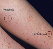
  • Pinpoint red spots on the skin, often found in groups that may look like a rash, caused by bleeding under the skin.These spots are called petechiae (see photo on the right).
    Petechiae-Purpura
    Petechiae & Purpura
  • Bruising or purplish areas (purpura) on the skin or mucous membranes caused by bleeding under the skin. (photo on the right)
  • More excessive bleeding can cause hematomas. A hematoma is a collection of clotted or partially clotted blood under the skin that feels like a lump. (photo lower right)
  • Nosebleeds, bleeding from the gums or excessive bleeding after injury.
  • Menstrual bleeding that is heavier than usual.
  • Some people experience untoward, otherwise unexplained fatigue when their platelet count is under 10,000/microl.
  • Hemorrhage is the most serious potential complication, intracranial (within the skull) being the most significant. (Note: the risk for major bleeding in otherwise healthy people is great only when the platelet count is less than 10,000/microl.)
    • Bleeding in the brain is rare. Symptoms of bleeding in the brain include:
      Sudden severe headaches, seizures with no previous history of seizures, weakness in arm(s) or leg(s), nausea or vomiting, decreased alertness, lethargy, changes in vision, tingling or numbness, difficulty speaking or understanding speech, difficulty swallowing, difficulty reading or writing, loss of fine motor skills (such as hand trembling), loss of coordination, loss of balance, abnormal sense of taste, or loss of consciousness. (Note: many of these symptoms are often caused by other conditions.)

    What can you do?
    Do some research if you are a considering HPV vaccination. Understand the potential risks associated with Gardasil and Cervarix as well as the potential benefits. Decide for yourself whether the potential benefits outweigh the potential risks. No one knows your family’s medical history better than you.
    If you have been vaccinated with either Gardasil or Cervarix, be aware that ITP is a possibility. Don’t panic, simply be vigilant. Talk to your medical provider if you are experiencing unusual bruising or unexplained rashes.
    If you are a medical professional, consider the possibility of ITP when examining patients who exhibit unexplained purpura after administration of HPV vaccines.
    If you are a medical consumer, contact the FDA and CDC. Ask them why such a high percentage of ITP reports in the VAERS database occurred after HPV vaccines. Ask them why two vaccines being associated with 18.5% of the ITP reports in VAERS does not raise a red flag. Ask them why they are not ordering studies to determine whether there could be a causal relationship between HPV vaccines and ITP.

Tuesday, April 23, 2013

Autism

Autism is a disorder of neural development characterized by impaired social interaction and communication, and by restricted and repetitive behavior. The diagnostic criteria require that symptoms become apparent before a child is three years old.[2] Autism affects information processing in the brain by altering how nerve cells and their synapses connect and organize; how this occurs is not well understood.[3] It is one of three recognized disorders in the autism spectrum (ASDs), the other two being Asperger syndrome, which lacks delays in cognitive development and language, and pervasive developmental disorder, not otherwise specified (commonly abbreviated as PDD-NOS), which is diagnosed when the full set of criteria for autism or Asperger syndrome are not met.[4]
Autism has a strong genetic basis, although the genetics of autism are complex and it is unclear whether ASD is explained more by rare mutations, or by rare combinations of common genetic variants.[5] In rare cases, autism is strongly associated with agents that cause birth defects.[6] Controversies surround other proposed environmental causes, such as heavy metals, pesticides or childhood vaccines;[7] the vaccine hypotheses are biologically implausible and lack convincing scientific evidence.[8] The prevalence of autism is about 1–2 per 1,000 people worldwide, and the Centers for Disease Control and Prevention (CDC) report 20 per 1,000 children in the United States are diagnosed with ASD as of 2012 (up from 11 per 1000 in 2008).[7][9][10] The number of people diagnosed with autism has been increasing dramatically since the 1980s, partly due to changes in diagnostic practice and government-subsidized financial incentives for named diagnoses;[10] the question of whether actual prevalence has increased is unresolved.[11]
Parents usually notice signs in the first two years of their child's life.[12] The signs usually develop gradually, but some autistic children first develop more normally and then regress.[13] Early behavioral or cognitive intervention can help autistic children gain self-care, social, and communication skills.[12] Although there is no known cure,[12] there have been reported cases of children who recovered.[14] Not many children with autism live independently after reaching adulthood, though some become successful.[15] An autistic culture has developed, with some individuals seeking a cure and others believing autism should be accepted as a difference and not treated as a disorder.[16]


Characteristics

Autism is a highly variable neurodevelopmental disorder[17] that first appears during infancy or childhood, and generally follows a steady course without remission.[18] Overt symptoms gradually begin after the age of six months, become established by age two or three years,[19] and tend to continue through adulthood, although often in more muted form.[20] It is distinguished not by a single symptom, but by a characteristic triad of symptoms: impairments in social interaction; impairments in communication; and restricted interests and repetitive behavior. Other aspects, such as atypical eating, are also common but are not essential for diagnosis.[21] Autism's individual symptoms occur in the general population and appear not to associate highly, without a sharp line separating pathologically severe from common traits.[22]

Social development

Social deficits distinguish autism and the related autism spectrum disorders (ASD; see Classification) from other developmental disorders.[20] People with autism have social impairments and often lack the intuition about others that many people take for granted. Noted autistic Temple Grandin described her inability to understand the social communication of neurotypicals, or people with normal neural development, as leaving her feeling "like an anthropologist on Mars".[23]
Unusual social development becomes apparent early in childhood. Autistic infants show less attention to social stimuli, smile and look at others less often, and respond less to their own name. Autistic toddlers differ more strikingly from social norms; for example, they have less eye contact and turn taking, and do not have the ability to use simple movements to express themselves, such as the deficiency to point at things.[24] Three- to five-year-old autistic children are less likely to exhibit social understanding, approach others spontaneously, imitate and respond to emotions, communicate nonverbally, and take turns with others. However, they do form attachments to their primary caregivers.[25] Most autistic children display moderately less attachment security than non-autistic children, although this difference disappears in children with higher mental development or less severe ASD.[26] Older children and adults with ASD perform worse on tests of face and emotion recognition.[27]
Children with high-functioning autism suffer from more intense and frequent loneliness compared to non-autistic peers, despite the common belief that children with autism prefer to be alone. Making and maintaining friendships often proves to be difficult for those with autism. For them, the quality of friendships, not the number of friends, predicts how lonely they feel. Functional friendships, such as those resulting in invitations to parties, may affect the quality of life more deeply.[28]
There are many anecdotal reports, but few systematic studies, of aggression and violence in individuals with ASD. The limited data suggest that, in children with mental retardation, autism is associated with aggression, destruction of property, and tantrums. A 2007 study interviewed parents of 67 children with ASD and reported that about two-thirds of the children had periods of severe tantrums and about one-third had a history of aggression, with tantrums significantly more common than in non-autistic children with language impairments.[29] A 2008 Swedish study found that, of individuals aged 15 or older discharged from hospital with a diagnosis of ASD, those who committed violent crimes were significantly more likely to have other psychopathological conditions such as psychosis.[30]

Communication

About a third to a half of individuals with autism do not develop enough natural speech to meet their daily communication needs.[31] Differences in communication may be present from the first year of life, and may include delayed onset of babbling, unusual gestures, diminished responsiveness, and vocal patterns that are not synchronized with the caregiver. In the second and third years, autistic children have less frequent and less diverse babbling, consonants, words, and word combinations; their gestures are less often integrated with words. Autistic children are less likely to make requests or share experiences, and are more likely to simply repeat others' words (echolalia)[32][33] or reverse pronouns.[34] Joint attention seems to be necessary for functional speech, and deficits in joint attention seem to distinguish infants with ASD:[4] for example, they may look at a pointing hand instead of the pointed-at object,[24][33] and they consistently fail to point at objects in order to comment on or share an experience.[4] Autistic children may have difficulty with imaginative play and with developing symbols into language.[32][33]
In a pair of studies, high-functioning autistic children aged 8–15 performed equally well as, and adults better than, individually matched controls at basic language tasks involving vocabulary and spelling. Both autistic groups performed worse than controls at complex language tasks such as figurative language, comprehension and inference. As people are often sized up initially from their basic language skills, these studies suggest that people speaking to autistic individuals are more likely to overestimate what their audience comprehends.[35]

Repetitive behavior

Autistic individuals display many forms of repetitive or restricted behavior, which the Repetitive Behavior Scale-Revised (RBS-R)[36] categorizes as follows.
Young boy asleep on a bed, facing the camera, with only the head visible and the body off-camera. On the bed behind the boy's head is a dozen or so toys carefully arranged in a line.
A young boy with autism who has arranged his toys in a row
  • Stereotypy is repetitive movement, such as hand flapping, head rolling, or body rocking.
  • Compulsive behavior is intended and appears to follow rules, such as arranging objects in stacks or lines.
  • Sameness is resistance to change; for example, insisting that the furniture not be moved or refusing to be interrupted.
  • Ritualistic behavior involves an unvarying pattern of daily activities, such as an unchanging menu or a dressing ritual. This is closely associated with sameness and an independent validation has suggested combining the two factors.[36]
  • Restricted behavior is limited in focus, interest, or activity, such as preoccupation with a single television program, toy, or game.
  • Self-injury includes movements that injure or can injure the person, such as eye poking, skin picking, hand biting, and head banging.[4] A 2007 study reported that self-injury at some point affected about 30% of children with ASD.[29]
No single repetitive or self-injurious behavior seems to be specific to autism, but only autism appears to have an elevated pattern of occurrence and severity of these behaviors.[37]

Other symptoms

Autistic individuals may have symptoms that are independent of the diagnosis, but that can affect the individual or the family.[21] An estimated 0.5% to 10% of individuals with ASD show unusual abilities, ranging from splinter skills such as the memorization of trivia to the extraordinarily rare talents of prodigious autistic savants.[38] Many individuals with ASD show superior skills in perception and attention, relative to the general population.[39] Sensory abnormalities are found in over 90% of those with autism, and are considered core features by some,[40] although there is no good evidence that sensory symptoms differentiate autism from other developmental disorders.[41] Differences are greater for under-responsivity (for example, walking into things) than for over-responsivity (for example, distress from loud noises) or for sensation seeking (for example, rhythmic movements).[42] An estimated 60%–80% of autistic people have motor signs that include poor muscle tone, poor motor planning, and toe walking;[40] deficits in motor coordination are pervasive across ASD and are greater in autism proper.[43]
Unusual eating behavior occurs in about three-quarters of children with ASD, to the extent that it was formerly a diagnostic indicator. Selectivity is the most common problem, although eating rituals and food refusal also occur;[29] this does not appear to result in malnutrition. Although some children with autism also have gastrointestinal (GI) symptoms, there is a lack of published rigorous data to support the theory that autistic children have more or different GI symptoms than usual;[44] studies report conflicting results, and the relationship between GI problems and ASD is unclear.[45]
Parents of children with ASD have higher levels of stress.[46] Siblings of children with ASD report greater admiration of and less conflict with the affected sibling than siblings of unaffected children and were similar to siblings of children with Down syndrome in these aspects of the sibling relationship. However, they reported lower levels of closeness and intimacy than siblings of children with Down syndrome; siblings of individuals with ASD have greater risk of negative well-being and poorer sibling relationships as adults.[47]

Classification

Autism is one of the five pervasive developmental disorders (PDD), which are characterized by widespread abnormalities of social interactions and communication, and severely restricted interests and highly repetitive behavior.[18] These symptoms do not imply sickness, fragility, or emotional disturbance.[20]
Of the five PDD forms, Asperger syndrome is closest to autism in signs and likely causes; Rett syndrome and childhood disintegrative disorder share several signs with autism, but may have unrelated causes; PDD not otherwise specified (PDD-NOS; also called atypical autism) is diagnosed when the criteria are not met for a more specific disorder.[48] Unlike with autism, people with Asperger syndrome have no substantial delay in language development.[2] The terminology of autism can be bewildering, with autism, Asperger syndrome and PDD-NOS often called the autism spectrum disorders (ASD)[12] or sometimes the autistic disorders,[49] whereas autism itself is often called autistic disorder, childhood autism, or infantile autism. In this article, autism refers to the classic autistic disorder; in clinical practice, though, autism, ASD, and PDD are often used interchangeably.[1] ASD, in turn, is a subset of the broader autism phenotype, which describes individuals who may not have ASD but do have autistic-like traits, such as avoiding eye contact.[50]
The manifestations of autism cover a wide spectrum, ranging from individuals with severe impairments—who may be silent, mentally disabled, and locked into hand flapping and rocking—to high functioning individuals who may have active but distinctly odd social approaches, narrowly focused interests, and verbose, pedantic communication.[51] Because the behavior spectrum is continuous, boundaries between diagnostic categories are necessarily somewhat arbitrary.[40] Sometimes the syndrome is divided into low-, medium- or high-functioning autism (LFA, MFA, and HFA), based on IQ thresholds,[52] or on how much support the individual requires in daily life; these subdivisions are not standardized and are controversial. Autism can also be divided into syndromal and non-syndromal autism; the syndromal autism is associated with severe or profound mental retardation or a congenital syndrome with physical symptoms, such as tuberous sclerosis.[53] Although individuals with Asperger syndrome tend to perform better cognitively than those with autism, the extent of the overlap between Asperger syndrome, HFA, and non-syndromal autism is unclear.[54]
Some studies have reported diagnoses of autism in children due to a loss of language or social skills, as opposed to a failure to make progress, typically from 15 to 30 months of age. The validity of this distinction remains controversial; it is possible that regressive autism is a specific subtype,[13][24][32][55] or that there is a continuum of behaviors between autism with and without regression.[56]
Research into causes has been hampered by the inability to identify biologically meaningful subgroups within the autistic population[57] and by the traditional boundaries between the disciplines of psychiatry, psychology, neurology and pediatrics.[58] Newer technologies such as fMRI and diffusion tensor imaging can help identify biologically relevant phenotypes (observable traits) that can be viewed on brain scans, to help further neurogenetic studies of autism;[59] one example is lowered activity in the fusiform face area of the brain, which is associated with impaired perception of people versus objects.[3] It has been proposed to classify autism using genetics as well as behavior.[60]

Causes

It has long been presumed that there is a common cause at the genetic, cognitive, and neural levels for autism's characteristic triad of symptoms.[61] However, there is increasing suspicion that autism is instead a complex disorder whose core aspects have distinct causes that often co-occur.[61][62]
Three diagrams of chromosome pairs A, B that are nearly identical. 1: B is missing a segment of A. 2: B has two adjacent copies of a segment of A. 3: B's copy of A's segment is in reverse order.
Deletion (1), duplication (2) and inversion (3) are all chromosome abnormalities that have been implicated in autism.[63]
Autism has a strong genetic basis, although the genetics of autism are complex and it is unclear whether ASD is explained more by rare mutations with major effects, or by rare multigene interactions of common genetic variants.[5][64] Complexity arises due to interactions among multiple genes, the environment, and epigenetic factors which do not change DNA but are heritable and influence gene expression.[20] Studies of twins suggest that heritability is 0.7 for autism and as high as 0.9 for ASD, and siblings of those with autism are about 25 times more likely to be autistic than the general population.[40] However, most of the mutations that increase autism risk have not been identified. Typically, autism cannot be traced to a Mendelian (single-gene) mutation or to a single chromosome abnormality, and none of the genetic syndromes associated with ASDs have been shown to selectively cause ASD.[5] Numerous candidate genes have been located, with only small effects attributable to any particular gene.[5] The large number of autistic individuals with unaffected family members may result from copy number variations—spontaneous deletions or duplications in genetic material during meiosis.[65] Hence, a substantial fraction of autism cases may be traceable to genetic causes that are highly heritable but not inherited: that is, the mutation that causes the autism is not present in the parental genome.[63]
Several lines of evidence point to synaptic dysfunction as a cause of autism.[3] Some rare mutations may lead to autism by disrupting some synaptic pathways, such as those involved with cell adhesion.[66] Gene replacement studies in mice suggest that autistic symptoms are closely related to later developmental steps that depend on activity in synapses and on activity-dependent changes.[67] All known teratogens (agents that cause birth defects) related to the risk of autism appear to act during the first eight weeks from conception, and though this does not exclude the possibility that autism can be initiated or affected later, it is strong evidence that autism arises very early in development.[6]
Although evidence for other environmental causes is anecdotal and has not been confirmed by reliable studies,[7] extensive searches are underway.[68] Environmental factors that have been claimed to contribute to or exacerbate autism, or may be important in future research, include certain foods, infectious disease, heavy metals, solvents, diesel exhaust, PCBs, phthalates and phenols used in plastic products, pesticides, brominated flame retardants, alcohol, smoking, illicit drugs, vaccines,[11] and prenatal stress,[69] although no links have been found, and some have been completely disproven.
Parents may first become aware of autistic symptoms in their child around the time of a routine vaccination. This has led to unsupported theories blaming vaccine "overload", a vaccine preservative, or the MMR vaccine for causing autism.[8] The latter theory was supported by a litigation-funded study that has since been shown to have been "an elaborate fraud".[70] Although these theories lack convincing scientific evidence and are biologically implausible,[8] parental concern about a potential vaccine link with autism has led to lower rates of childhood immunizations, outbreaks of previously controlled childhood diseases in some countries, and the preventable deaths of several children.[71][72]

Mechanism

Autism's symptoms result from maturation-related changes in various systems of the brain. How autism occurs is not well understood. Its mechanism can be divided into two areas: the pathophysiology of brain structures and processes associated with autism, and the neuropsychological linkages between brain structures and behaviors.[73] The behaviors appear to have multiple pathophysiologies.[22]

Pathophysiology

Two diagrams of major brain structures implicated in autism. The upper diagram shows the cerebral cortex near the top and the basal ganglia in the center, just above the amygdala and hippocampus. The lower diagram shows the corpus callosum near the center, the cerebellum in the lower rear, and the brain stem in the lower center.
Autism affects the amygdala, cerebellum, and many other parts of the brain.[74]
Unlike many other brain disorders, such as Parkinson's, autism does not have a clear unifying mechanism at either the molecular, cellular, or systems level; it is not known whether autism is a few disorders caused by mutations converging on a few common molecular pathways, or is (like intellectual disability) a large set of disorders with diverse mechanisms.[17] Autism appears to result from developmental factors that affect many or all functional brain systems,[75] and to disturb the timing of brain development more than the final product.[74] Neuroanatomical studies and the associations with teratogens strongly suggest that autism's mechanism includes alteration of brain development soon after conception.[6] This anomaly appears to start a cascade of pathological events in the brain that are significantly influenced by environmental factors.[76] Just after birth, the brains of autistic children tend to grow faster than usual, followed by normal or relatively slower growth in childhood. It is not known whether early overgrowth occurs in all autistic children. It seems to be most prominent in brain areas underlying the development of higher cognitive specialization.[40] Hypotheses for the cellular and molecular bases of pathological early overgrowth include the following:
Interactions between the immune system and the nervous system begin early during the embryonic stage of life, and successful neurodevelopment depends on a balanced immune response. Aberrant immune activity during critical periods of neurodevelopment is possibly part of the mechanism of some forms of ASD.[84] Although some abnormalities in the immune system have been found in specific subgroups of autistic individuals, it is not known whether these abnormalities are relevant to or secondary to autism's disease processes.[85] As autoantibodies are found in conditions other than ASD, and are not always present in ASD,[86] the relationship between immune disturbances and autism remains unclear and controversial.[78]
The relationship of neurochemicals to autism is not well understood; several have been investigated, with the most evidence for the role of serotonin and of genetic differences in its transport.[3] The role of group I metabotropic glutamate receptors (mGluR) in the pathogenesis of fragile X syndrome, the most common identified genetic cause of autism, has led to interest in the possible implications for future autism research into this pathway.[87] Some data suggest an increase in several growth hormones; other data argue for diminished growth factors.[88] Also, some inborn errors of metabolism are associated with autism, but probably account for less than 5% of cases.[89]
The mirror neuron system (MNS) theory of autism hypothesizes that distortion in the development of the MNS interferes with imitation and leads to autism's core features of social impairment and communication difficulties. The MNS operates when an animal performs an action or observes another animal perform the same action. The MNS may contribute to an individual's understanding of other people by enabling the modeling of their behavior via embodied simulation of their actions, intentions, and emotions.[90] Several studies have tested this hypothesis by demonstrating structural abnormalities in MNS regions of individuals with ASD, delay in the activation in the core circuit for imitation in individuals with Asperger syndrome, and a correlation between reduced MNS activity and severity of the syndrome in children with ASD.[91] However, individuals with autism also have abnormal brain activation in many circuits outside the MNS[92] and the MNS theory does not explain the normal performance of autistic children on imitation tasks that involve a goal or object.[93]
A human brain viewed from above. About 10% is highlighted in yellow and 10% in blue. There is only a tiny (perhaps 0.5%) green region where they overlap.
Autistic individuals tend to use different areas of the brain (yellow) for a movement task compared to a control group (blue).[94]
ASD-related patterns of low function and aberrant activation in the brain differ depending on whether the brain is doing social or nonsocial tasks.[95] In autism there is evidence for reduced functional connectivity of the default network, a large-scale brain network involved in social and emotional processing, with intact connectivity of the task-positive network, used in sustained attention and goal-directed thinking. In people with autism the two networks are not negatively correlated in time, suggesting an imbalance in toggling between the two networks, possibly reflecting a disturbance of self-referential thought.[96] A 2008 brain-imaging study found a specific pattern of signals in the cingulate cortex which differs in individuals with ASD.[97]
The underconnectivity theory of autism hypothesizes that autism is marked by underfunctioning high-level neural connections and synchronization, along with an excess of low-level processes.[98] Evidence for this theory has been found in functional neuroimaging studies on autistic individuals[35] and by a brainwave study that suggested that adults with ASD have local overconnectivity in the cortex and weak functional connections between the frontal lobe and the rest of the cortex.[99] Other evidence suggests the underconnectivity is mainly within each hemisphere of the cortex and that autism is a disorder of the association cortex.[100]
From studies based on event-related potentials, transient changes to the brain's electrical activity in response to stimuli, there is considerable evidence for differences in autistic individuals with respect to attention, orientiation to auditory and visual stimuli, novelty detection, language and face processing, and information storage; several studies have found a preference for nonsocial stimuli.[101] For example, magnetoencephalography studies have found evidence in autistic children of delayed responses in the brain's processing of auditory signals.[102]
In the genetic area, relations have been found between autism and schizophrenia based on duplications and deletions of chromosomes; research showed that schizophrenia and autism are significantly more common in combination with 1q21.1 deletion syndrome. Research on autism/schizophrenia relations for chromosome 15 (15q13.3), chromosome 16 (16p13.1) and chromosome 17 (17p12) are inconclusive.[103]

Neuropsychology

Two major categories of cognitive theories have been proposed about the links between autistic brains and behavior.
The first category focuses on deficits in social cognition. The empathizing–systemizing theory postulates that autistic individuals can systemize—that is, they can develop internal rules of operation to handle events inside the brain—but are less effective at empathizing by handling events generated by other agents. An extension, the extreme male brain theory, hypothesizes that autism is an extreme case of the male brain, defined psychometrically as individuals in whom systemizing is better than empathizing;[104] this extension is controversial, as many studies contradict the idea that baby boys and girls respond differently to people and objects.[105]
These theories are somewhat related to the earlier theory of mind approach, which hypothesizes that autistic behavior arises from an inability to ascribe mental states to oneself and others. The theory of mind hypothesis is supported by autistic children's atypical responses to the Sally–Anne test for reasoning about others' motivations,[104] and the mirror neuron system theory of autism described in Pathophysiology maps well to the hypothesis.[91] However, most studies have found no evidence of impairment in autistic individuals' ability to understand other people's basic intentions or goals; instead, data suggests that impairments are found in understanding more complex social emotions or in considering others' viewpoints.[106]
The second category focuses on nonsocial or general processing: the executive functions such as working memory, planning, inhibition. In his review, Kenworthy states that "the claim of executive dysfunction as a causal factor in autism is controversial", however, "it is clear that executive dysfunction plays a role in the social and cognitive deficits observed in individuals with autism".[107] Tests of core executive processes such as eye movement tasks indicate improvement from late childhood to adolescence, but performance never reaches typical adult levels.[108] A strength of the theory is predicting stereotyped behavior and narrow interests;[109] two weaknesses are that executive function is hard to measure[107] and that executive function deficits have not been found in young autistic children.[27]
Weak central coherence theory hypothesizes that a limited ability to see the big picture underlies the central disturbance in autism. One strength of this theory is predicting special talents and peaks in performance in autistic people.[110] A related theory—enhanced perceptual functioning—focuses more on the superiority of locally oriented and perceptual operations in autistic individuals.[111] These theories map well from the underconnectivity theory of autism.
Neither category is satisfactory on its own; social cognition theories poorly address autism's rigid and repetitive behaviors, while the nonsocial theories have difficulty explaining social impairment and communication difficulties.[62] A combined theory based on multiple deficits may prove to be more useful.[112]

Screening

About half of parents of children with ASD notice their child's unusual behaviors by age 18 months, and about four-fifths notice by age 24 months.[55] According to an article in the Journal of Autism and Developmental Disorders, failure to meet any of the following milestones "is an absolute indication to proceed with further evaluations. Delay in referral for such testing may delay early diagnosis and treatment and affect the long-term outcome".[21]
  • No babbling by 12 months.
  • No gesturing (pointing, waving, etc.) by 12 months.
  • No single words by 16 months.
  • No two-word (spontaneous, not just echolalic) phrases by 24 months.
  • Any loss of any language or social skills, at any age.
US and Japanese practice is to screen all children for ASD at 18 and 24 months, using autism-specific formal screening tests. In contrast, in the UK, children whose families or doctors recognize possible signs of autism are screened. It is not known which approach is more effective.[3] Screening tools include the Modified Checklist for Autism in Toddlers (M-CHAT), the Early Screening of Autistic Traits Questionnaire, and the First Year Inventory; initial data on M-CHAT and its predecessor CHAT on children aged 18–30 months suggests that it is best used in a clinical setting and that it has low sensitivity (many false-negatives) but good specificity (few false-positives).[55] It may be more accurate to precede these tests with a broadband screener that does not distinguish ASD from other developmental disorders.[113] Screening tools designed for one culture's norms for behaviors like eye contact may be inappropriate for a different culture.[114] Although genetic screening for autism is generally still impractical, it can be considered in some cases, such as children with neurological symptoms and dysmorphic features.[115]

Diagnosis

Diagnosis is based on behavior, not cause or mechanism.[22][116] Autism is defined in the DSM-IV-TR as exhibiting at least six symptoms total, including at least two symptoms of qualitative impairment in social interaction, at least one symptom of qualitative impairment in communication, and at least one symptom of restricted and repetitive behavior. Sample symptoms include lack of social or emotional reciprocity, stereotyped and repetitive use of language or idiosyncratic language, and persistent preoccupation with parts of objects. Onset must be prior to age three years, with delays or abnormal functioning in either social interaction, language as used in social communication, or symbolic or imaginative play. The disturbance must not be better accounted for by Rett syndrome or childhood disintegrative disorder.[2] ICD-10 uses essentially the same definition.[18]
Several diagnostic instruments are available. Two are commonly used in autism research: the Autism Diagnostic Interview-Revised (ADI-R) is a semistructured parent interview, and the Autism Diagnostic Observation Schedule (ADOS)[117] uses observation and interaction with the child. The Childhood Autism Rating Scale (CARS) is used widely in clinical environments to assess severity of autism based on observation of children.[24]
A pediatrician commonly performs a preliminary investigation by taking developmental history and physically examining the child. If warranted, diagnosis and evaluations are conducted with help from ASD specialists, observing and assessing cognitive, communication, family, and other factors using standardized tools, and taking into account any associated medical conditions.[118] A pediatric neuropsychologist is often asked to assess behavior and cognitive skills, both to aid diagnosis and to help recommend educational interventions.[119] A differential diagnosis for ASD at this stage might also consider mental retardation, hearing impairment, and a specific language impairment[118] such as Landau–Kleffner syndrome.[120] The presence of autism can make it harder to diagnose coexisting psychiatric disorders such as depression.[121]
Clinical genetics evaluations are often done once ASD is diagnosed, particularly when other symptoms already suggest a genetic cause.[1] Although genetic technology allows clinical geneticists to link an estimated 40% of cases to genetic causes,[122] consensus guidelines in the US and UK are limited to high-resolution chromosome and fragile X testing.[1] A genotype-first model of diagnosis has been proposed, which would routinely assess the genome's copy number variations.[123] As new genetic tests are developed several ethical, legal, and social issues will emerge. Commercial availability of tests may precede adequate understanding of how to use test results, given the complexity of autism's genetics.[124] Metabolic and neuroimaging tests are sometimes helpful, but are not routine.[1]
ASD can sometimes be diagnosed by age 14 months, although diagnosis becomes increasingly stable over the first three years of life: for example, a one-year-old who meets diagnostic criteria for ASD is less likely than a three-year-old to continue to do so a few years later.[55] In the UK the National Autism Plan for Children recommends at most 30 weeks from first concern to completed diagnosis and assessment, though few cases are handled that quickly in practice.[118] A 2009 US study found the average age of formal ASD diagnosis was 5.7 years, far above recommendations, and that 27% of children remained undiagnosed at age 8 years.[125] Although the symptoms of autism and ASD begin early in childhood, they are sometimes missed; years later, adults may seek diagnoses to help them or their friends and family understand themselves, to help their employers make adjustments, or in some locations to claim disability living allowances or other benefits.[126]
Underdiagnosis and overdiagnosis are problems in marginal cases, and much of the recent increase in the number of reported ASD cases is likely due to changes in diagnostic practices. The increasing popularity of drug treatment options and the expansion of benefits has given providers incentives to diagnose ASD, resulting in some overdiagnosis of children with uncertain symptoms. Conversely, the cost of screening and diagnosis and the challenge of obtaining payment can inhibit or delay diagnosis.[127] It is particularly hard to diagnose autism among the visually impaired, partly because some of its diagnostic criteria depend on vision, and partly because autistic symptoms overlap with those of common blindness syndromes or blindisms.[128]

Management

A young child points, in front of a woman who smiles and points in the same direction.
A three-year-old with autism points to fish in an aquarium, as part of an experiment on the effect of intensive shared-attention training on language development.[94]
The main goals when treating children with autism are to lessen associated deficits and family distress, and to increase quality of life and functional independence. No single treatment is best and treatment is typically tailored to the child's needs.[12] Families and the educational system are the main resources for treatment.[3] Studies of interventions have methodological problems that prevent definitive conclusions about efficacy.[129] Although many psychosocial interventions have some positive evidence, suggesting that some form of treatment is preferable to no treatment, the methodological quality of systematic reviews of these studies has generally been poor, their clinical results are mostly tentative, and there is little evidence for the relative effectiveness of treatment options.[130] Intensive, sustained special education programs and behavior therapy early in life can help children acquire self-care, social, and job skills,[12] and often improve functioning and decrease symptom severity and maladaptive behaviors;[131] claims that intervention by around age three years is crucial are not substantiated.[132] Available approaches include applied behavior analysis (ABA), developmental models, structured teaching, speech and language therapy, social skills therapy, and occupational therapy.[12]
Educational interventions can be effective to varying degrees in most children: intensive ABA treatment has demonstrated effectiveness in enhancing global functioning in preschool children[133] and is well-established for improving intellectual performance of young children.[131] Neuropsychological reports are often poorly communicated to educators, resulting in a gap between what a report recommends and what education is provided.[119] It is not known whether treatment programs for children lead to significant improvements after the children grow up,[131] and the limited research on the effectiveness of adult residential programs shows mixed results.[134] The appropriateness of including children with varying severity of autism spectrum disorders in the general education population is a subject of current debate among educators and researchers.[135]
Many medications are used to treat ASD symptoms that interfere with integrating a child into home or school when behavioral treatment fails.[20][136] More than half of US children diagnosed with ASD are prescribed psychoactive drugs or anticonvulsants, with the most common drug classes being antidepressants, stimulants, and antipsychotics.[137] Aside from antipsychotics,[138] there is scant reliable research about the effectiveness or safety of drug treatments for adolescents and adults with ASD.[139] A person with ASD may respond atypically to medications, the medications can have adverse effects,[12] and no known medication relieves autism's core symptoms of social and communication impairments.[140] Experiments in mice have reversed or reduced some symptoms related to autism by replacing or modulating gene function,[67][87] suggesting the possibility of targeting therapies to specific rare mutations known to cause autism.[66][141]
Although many alternative therapies and interventions are available, few are supported by scientific studies.[27][142] Treatment approaches have little empirical support in quality-of-life contexts, and many programs focus on success measures that lack predictive validity and real-world relevance.[28] Scientific evidence appears to matter less to service providers than program marketing, training availability, and parent requests.[143] Some alternative treatments may place the child at risk. A 2008 study found that compared to their peers, autistic boys have significantly thinner bones if on casein-free diets;[144] in 2005, botched chelation therapy killed a five-year-old child with autism.[145]
Treatment is expensive; indirect costs are more so. For someone born in 2000, a US study estimated an average lifetime cost of $3.94 million (net present value in 2013 dollars, inflation-adjusted from 2003 estimate),[146] with about 10% medical care, 30% extra education and other care, and 60% lost economic productivity.[147] Publicly supported programs are often inadequate or inappropriate for a given child, and unreimbursed out-of-pocket medical or therapy expenses are associated with likelihood of family financial problems;[148] one 2008 US study found a 14% average loss of annual income in families of children with ASD,[149] and a related study found that ASD is associated with higher probability that child care problems will greatly affect parental employment.[150] US states increasingly require private health insurance to cover autism services, shifting costs from publicly funded education programs to privately funded health insurance.[151] After childhood, key treatment issues include residential care, job training and placement, sexuality, social skills, and estate planning.[152]

Prognosis

There is no known cure.[3][12] Children recover occasionally, so that they lose their diagnosis of ASD;[14] this occurs sometimes after intensive treatment and sometimes not. It is not known how often recovery happens;[131] reported rates in unselected samples of children with ASD have ranged from 3% to 25%.[14] Most autistic children can acquire language by age 5 or younger, though a few have developed communication skills in later years.[153] Most children with autism lack social support, meaningful relationships, future employment opportunities or self-determination.[28] Although core difficulties tend to persist, symptoms often become less severe with age.[20]
Few high-quality studies address long-term prognosis. Some adults show modest improvement in communication skills, but a few decline; no study has focused on autism after midlife.[154] Acquiring language before age six, having an IQ above 50, and having a marketable skill all predict better outcomes; independent living is unlikely with severe autism.[155] A 2004 British study of 68 adults who were diagnosed before 1980 as autistic children with IQ above 50 found that 12% achieved a high level of independence as adults, 10% had some friends and were generally in work but required some support, 19% had some independence but were generally living at home and needed considerable support and supervision in daily living, 46% needed specialist residential provision from facilities specializing in ASD with a high level of support and very limited autonomy, and 12% needed high-level hospital care.[15] A 2005 Swedish study of 78 adults that did not exclude low IQ found worse prognosis; for example, only 4% achieved independence.[156] A 2008 Canadian study of 48 young adults diagnosed with ASD as preschoolers found outcomes ranging through poor (46%), fair (32%), good (17%), and very good (4%); 56% of these young adults had been employed at some point during their lives, mostly in volunteer, sheltered or part-time work.[157] Changes in diagnostic practice and increased availability of effective early intervention make it unclear whether these findings can be generalized to recently diagnosed children.[11]

Epidemiology

Bar chart versus time. The graph rises steadily from 1996 to 2007, from about 0.7 to about 5.3. The trend curves slightly upward.
Reports of autism cases per 1,000 children grew dramatically in the US from 1996 to 2007. It is unknown how much, if any, growth came from changes in autism's prevalence.
Most recent reviews tend to estimate a prevalence of 1–2 per 1,000 for autism and close to 6 per 1,000 for ASD,[11] and 11 per 1,000 children in the United States for ASD as of 2008;[9][158] because of inadequate data, these numbers may underestimate ASD's true prevalence.[1] PDD-NOS's prevalence has been estimated at 3.7 per 1,000, Asperger syndrome at roughly 0.6 per 1,000, and childhood disintegrative disorder at 0.02 per 1,000.[159] The number of reported cases of autism increased dramatically in the 1990s and early 2000s. This increase is largely attributable to changes in diagnostic practices, referral patterns, availability of services, age at diagnosis, and public awareness,[159][160] though unidentified environmental risk factors cannot be ruled out.[7] The available evidence does not rule out the possibility that autism's true prevalence has increased;[159] a real increase would suggest directing more attention and funding toward changing environmental factors instead of continuing to focus on genetics.[68]
Boys are at higher risk for ASD than girls. The sex ratio averages 4.3:1 and is greatly modified by cognitive impairment: it may be close to 2:1 with mental retardation and more than 5.5:1 without.[11] Although the evidence does not implicate any single pregnancy-related risk factor as a cause of autism, the risk of autism is associated with advanced age in either parent, and with diabetes, bleeding, and use of psychiatric drugs in the mother during pregnancy.[161] The risk is greater with older fathers than with older mothers; two potential explanations are the known increase in mutation burden in older sperm, and the hypothesis that men marry later if they carry genetic liability and show some signs of autism.[40] Most professionals believe that race, ethnicity, and socioeconomic background do not affect the occurrence of autism.[162]
Several other conditions are common in children with autism.[3] They include:
  • Genetic disorders. About 10–15% of autism cases have an identifiable Mendelian (single-gene) condition, chromosome abnormality, or other genetic syndrome,[163] and ASD is associated with several genetic disorders.[164]
  • Mental retardation. The percentage of autistic individuals who also meet criteria for mental retardation has been reported as anywhere from 25% to 70%, a wide variation illustrating the difficulty of assessing autistic intelligence.[165] In comparison, for PDD-NOS the association with mental retardation is much weaker,[166] and by definition, the diagnosis of Asperger's excludes mental retardation.[167]
  • Anxiety disorders are common among children with ASD; there are no firm data, but studies have reported prevalences ranging from 11% to 84%. Many anxiety disorders have symptoms that are better explained by ASD itself, or are hard to distinguish from ASD's symptoms.[168]
  • Epilepsy, with variations in risk of epilepsy due to age, cognitive level, and type of language disorder.[169]
  • Several metabolic defects, such as phenylketonuria, are associated with autistic symptoms.[89]
  • Minor physical anomalies are significantly increased in the autistic population.[170]
  • Preempted diagnoses. Although the DSM-IV rules out concurrent diagnosis of many other conditions along with autism, the full criteria for Attention deficit hyperactivity disorder (ADHD), Tourette syndrome, and other of these conditions are often present and these comorbid diagnoses are increasingly accepted.[171]
  • Sleep problems affect about two-thirds of individuals with ASD at some point in childhood. These most commonly include symptoms of insomnia such as difficulty in falling asleep, frequent nocturnal awakenings, and early morning awakenings. Sleep problems are associated with difficult behaviors and family stress, and are often a focus of clinical attention over and above the primary ASD diagnosis.[172]

History

Head and shoulders of a man in his early 60s in coat and tie, facing slightly to his right. He is balding and has a serious but slightly smiling expression.
Leo Kanner introduced the label early infantile autism in 1943.
A few examples of autistic symptoms and treatments were described long before autism was named. The Table Talk of Martin Luther, compiled by his notetaker, Mathesius, contains the story of a 12-year-old boy who may have been severely autistic.[173] Luther reportedly thought the boy was a soulless mass of flesh possessed by the devil, and suggested that he be suffocated, although a later critic has cast doubt on the veracity of this report.[174] The earliest well-documented case of autism is that of Hugh Blair of Borgue, as detailed in a 1747 court case in which his brother successfully petitioned to annul Blair's marriage to gain Blair's inheritance.[175] The Wild Boy of Aveyron, a feral child caught in 1798, showed several signs of autism; the medical student Jean Itard treated him with a behavioral program designed to help him form social attachments and to induce speech via imitation.[176]
The New Latin word autismus (English translation autism) was coined by the Swiss psychiatrist Eugen Bleuler in 1910 as he was defining symptoms of schizophrenia. He derived it from the Greek word autós (αὐτός, meaning self), and used it to mean morbid self-admiration, referring to "autistic withdrawal of the patient to his fantasies, against which any influence from outside becomes an intolerable disturbance".[177]
The word autism first took its modern sense in 1938 when Hans Asperger of the Vienna University Hospital adopted Bleuler's terminology autistic psychopaths in a lecture in German about child psychology.[178] Asperger was investigating an ASD now known as Asperger syndrome, though for various reasons it was not widely recognized as a separate diagnosis until 1981.[176] Leo Kanner of the Johns Hopkins Hospital first used autism in its modern sense in English when he introduced the label early infantile autism in a 1943 report of 11 children with striking behavioral similarities.[34] Almost all the characteristics described in Kanner's first paper on the subject, notably "autistic aloneness" and "insistence on sameness", are still regarded as typical of the autistic spectrum of disorders.[62] It is not known whether Kanner derived the term independently of Asperger.[179]
Kanner's reuse of autism led to decades of confused terminology like infantile schizophrenia, and child psychiatry's focus on maternal deprivation led to misconceptions of autism as an infant's response to "refrigerator mothers". Starting in the late 1960s autism was established as a separate syndrome by demonstrating that it is lifelong, distinguishing it from mental retardation and schizophrenia and from other developmental disorders, and demonstrating the benefits of involving parents in active programs of therapy.[180] As late as the mid-1970s there was little evidence of a genetic role in autism; now it is thought to be one of the most heritable of all psychiatric conditions.[181] Although the rise of parent organizations and the destigmatization of childhood ASD have deeply affected how we view ASD,[176] parents continue to feel social stigma in situations where their autistic children's behaviors are perceived negatively by others,[182] and many primary care physicians and medical specialists still express some beliefs consistent with outdated autism research.[183]
The Internet has helped autistic individuals bypass nonverbal cues and emotional sharing that they find so hard to deal with, and has given them a way to form online communities and work remotely.[184] Sociological and cultural aspects of autism have developed: some in the community seek a cure, while others believe that autism is simply another way of being.[16][185]